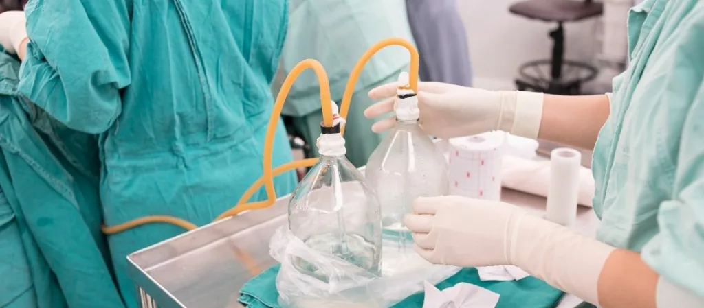
A drain is a medical device used after surgery to remove fluid accumulation from the operated area. By preventing hematoma or seroma formation, drains support proper wound healing and reduce the risk of infection.
Drain usage in plastic surgery is common, especially after procedures involving large tissue surfaces. Proper placement and monitoring are crucial for maintaining safe and effective recovery.
How to care for a surgical drain involves regular emptying, measuring output, and keeping the entry site clean. Patients are advised to follow their surgeon’s instructions precisely to minimize complications.
The removal of drains depends on the volume and type of fluid collected. Typically, drains are removed once output decreases, ensuring the tissue has healed sufficiently to continue recovery without assistance.
What Is a Drain and Why Is It Important in Surgery?
A drain, simply put, is a “tube” or “conduit” system used after surgery or injury to expel unwanted fluids (blood, purulent discharge, lymph, etc.) from the body. Just as a kitchen sink has a pipe to prevent clogging, a surgical drain gives excess fluid in wounded tissues an “exit route” so it can “flow away.”
Post-operative accumulations of blood (hematoma) or tissue fluid (seroma) can prolong healing and raise infection risk. Surgeons therefore insert drains after certain procedures to keep control. From large abdominal operations to plastic-surgery procedures such as breast reconstruction, drains are widely used. These small yet effective devices remove the “unwanted pools” in the body, allowing tissues to fuse more quickly and healthily.
To see why drains matter, consider what happens when excess fluid collects at a wound or incision. Blood or serous fluids offer an ideal breeding ground for microbes. They can also separate tissue layers, hampering wound edges from knitting together. Here lies the drain’s value: it regularly removes fluids that would otherwise hinder tissue adhesion or invite bacterial growth, keeping the surgical site clean and speeding recovery.
What Are the Types of Drains?
Drains are medical devices used during surgical procedures to remove fluids (such as blood, pus, or lymph) from the body.
Different types of drains have been developed to meet various needs:
- Passive Drains: Fluids exit the body through gravity and capillary action (e.g., Penrose drain).
- Active Drains: Use negative pressure (vacuum) to remove fluids (e.g., Hemovac, Jackson-Pratt drain).
- Open Drains: Fluid flows directly out of the body, posing a higher risk of infection.
- Closed Drains: Fluid collects in a sterile reservoir, reducing the risk of infection.
- Subcutaneous Drains: Used to drain fluids from under the skin.
- Thoracostomy Tube: Used to remove air or fluid from the chest cavity.
How Does a Drain Work to Remove Fluids from the Body?
To grasp how drains function, divide them into two main categories: passive and active. Passive drains rely on gravity or simple capillarity (fluid seeping through a narrow channel). Old-style rubber or plastic ribbon drains (often called “Penrose drains”) direct fluid out of the wound via pressure differences without any external suction—like opening a window and allowing air to drift in.
Active drain systems work on negative pressure or suction. A classic example is the Jackson-Pratt (JP) drain. A JP consists of a thin tube placed in the surgical site and a soft “bulb” reservoir attached outside. When the bulb is squeezed and sealed, it creates a vacuum that draws fluid out of the wound—much like a household vacuum cleaner. By keeping the area dry, a JP drain prevents blood or lymph from collecting.
For a drain to work efficiently, the surgeon must position it correctly during the operation. If placed too far from the target area or incorrectly aligned, fluid may continue to pool. Tissue healing can also clog the drain, just as limescale blocks a sink pipe. Therefore, patients with drains may stay in hospital for observation or, if discharged, must follow care instructions (checking the line, emptying the bulb) carefully.
When Are Drains Typically Used After an Operation?
Drain usage has become nearly routine after certain surgeries. In orthopedic procedures—especially large joint replacements—drains prevent blood from pooling in the wound, which could otherwise foster bacteria. In abdominal surgery (bowel or stomach operations), drains help remove fluid or air from risky areas, easing recovery.
In breast surgery and abdominoplasty (tummy tuck), drains counter seroma formation. Because wide tissue dissection creates an under-skin pocket prone to lymph build-up, drains evacuate that “pocket,” much like emptying a space that tends to fill with water.
Yet drain use is not an absolute rule. Some surgeons employ drains only “when necessary,” while others may omit them altogether in minor cases. The decision depends on the patient’s condition, surgical technique, and the surgeon’s experience. In head-and-neck surgery, for instance, a small incision may not warrant a drain, whereas a broader dissection might. In short, drain placement reflects the surgeon’s assessment of potential complications.
What Different Types of Drains Can Be Used?
In practice, drains are classified as open or closed, passive or active, to suit varied surgical needs.
- Open Drains (Passive): Classic Penrose or corrugated rubber/plastic sheets belong here. Fluid exits by gravity or pressure difference and usually soaks into external gauze. They are simple and inexpensive but carry a higher infection risk due to their open nature.
- Closed Drains (Active or Passive): These feature a collection bag or bulb, isolating the system from the environment. Jackson-Pratt (JP) or Hemovac drains create negative pressure within a reservoir to actively pull fluid. Their closed design reduces microbial entry.
- Mini-Vacuum Drains: Used in smaller surgical fields expecting minimal fluid. They also use suction but have limited capacity.
- Redivac Drains: Provide high suction for large surgeries needing powerful fluid evacuation. Their strong vacuum helps prevent significant hematoma or seroma.
- Pigtail Drains: Employed in deeper cavities like the abdomen or chest where fluid is localized. The curled tip anchors within tissue and drains a broad area.
- Thoracostomy Tube (Chest Tube): Removes air or fluid from the pleural space, restoring negative intrathoracic pressure—vital for lung re-expansion after collapse or effusion.
Choosing the right drain, like selecting the correct screwdriver tip, greatly aids recovery.
How Is a Drain Placed into the Body?
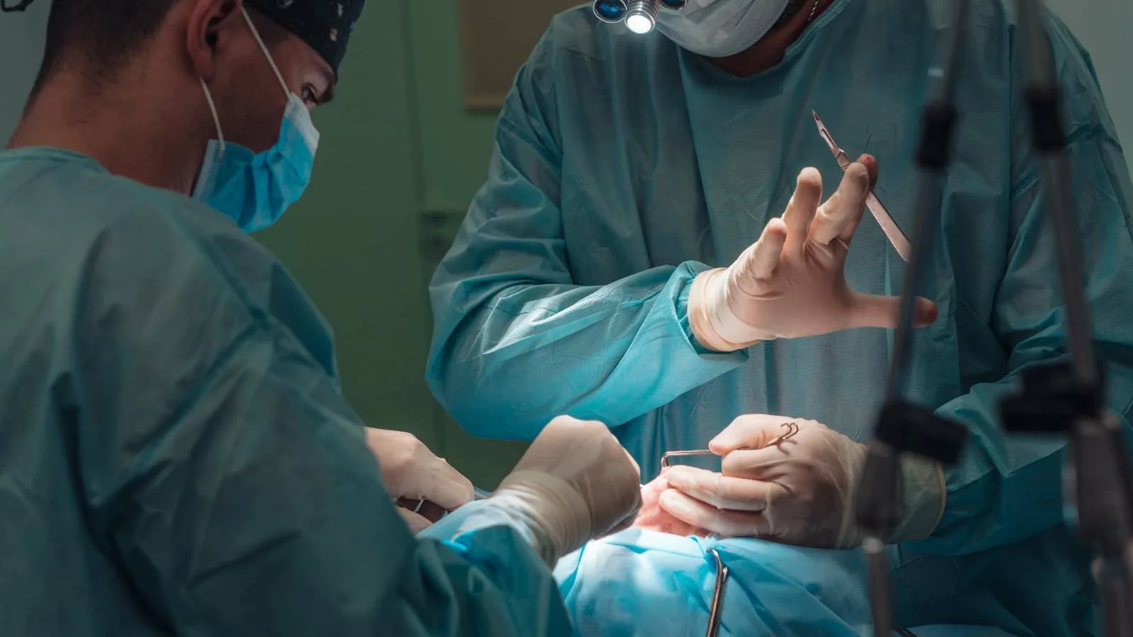
Drain insertion is a deliberate step in surgery. Near the end of the procedure—or during pre-operative planning—the surgeon selects the optimal site, sometimes aided by ultrasound or CT imaging.
Technically, a drain is usually a thin, flexible tube. The surgeon creates a small skin incision or stab wound and threads the tube to the surgical area—occasionally using a guide called a trocar. After ensuring the tip lies at the intended spot, the tube exits through the skin and is secured with sutures or fixation devices, much like buttons keep clothing in place. A dressing or adhesive protects the entry site. If it is an active system (e.g., JP drain), a bulb or reservoir is attached externally and begins suction. With passive drains, fluid may drip into a bag or onto gauze.
The team then checks for leaks, kinks, or obstructions. Once everything works, the surgical wound is closed and the patient awakened. Drain placement is complete.
What Are the Benefits of Using a Drain After Surgery?
Drains provide several advantages post-operatively. The foremost is reduced infection risk. Excess fluid acts as “nutritious soup” for bacteria; draining it denies microbes that environment. Blood (hematoma) or lymph (seroma) collections are prime microbial habitats; quick removal curbs colonization.
Drains also lessen pain and swelling. Fluid under tension presses on tissues, causing discomfort. Draining reduces pressure, improving patient comfort and mobility—especially important in orthopedic cases requiring early physiotherapy.
Furthermore, drains speed tissue healing. Two surfaces fuse best without fluid between them—like two sheets of paper sticking only if they’re dry. Continuous drainage clears the gap, letting tissues “glue” together.
Finally, drains help detect complications early. A sudden change in the color or volume of drainage after abdominal surgery, for example, may signal leakage or bleeding, prompting swift intervention.
What Are the Possible Complications of Using a Drain?
As with any medical practice, drains carry risks. Knowing and preventing them is vital.
- Infection: A drain links internal tissue to the outside world, a pathway for microbes. Redness, discharge, or fever at the entry site demands prompt care—antibiotics or drain replacement.
- Pain and Discomfort: Patients may feel soreness, stinging, or friction. Proper dressing and secure fixation ease these symptoms.
- Blockage or Breakage: Clots, tissue debris, or poor maintenance can clog the line, preventing drainage. Rarely, the tube may fracture, leaving a piece inside, requiring an additional procedure.
- Tissue Damage or Misplacement: An incorrectly positioned drain can harm healthy tissue, particularly in sensitive areas (joints, brain, thorax). Careful placement is essential.
- Persistent Seroma or Hematoma: Fluid may still collect if the drain clogs or care is insufficient. Early recognition allows flushing or replacement to restore flow.
Expert insertion, regular dressing, hygiene, and adherence to instructions minimize these risks.
How Long Does a Drain Stay in Place After Surgery?
Duration depends on surgery type, patient condition, and daily drainage volume. Some drains remain only a few days; others stay weeks. Where risk is high, longer use may occur.
After abdominal surgery, the drain may stay until daily output falls below a threshold (e.g., 20-30 ml/day) for several consecutive days. In head-and-neck procedures, drains are often removed sooner, as fluid accumulation is minimal. Joint surgeries may need two-three days; major spinal operations five-six. In breast or tummy-tuck procedures, drains can remain one-two weeks or more, varying by individual healing.
Because infection risk rises with prolonged placement, doctors aim for “the shortest duration that achieves maximum benefit.” Daily recording of output guides timing of removal.
How Should Patients Care for a Drain at Home?
With shorter hospital stays, many patients go home with drains. Basic knowledge is essential.
First rule: cleanliness. Change the dressing at least once daily—or as instructed. Wash hands thoroughly and disinfect them. Gently clean drainage residue at the entry site using clean gauze, then cover with the recommended dressing.
For an active drain (e.g., Jackson-Pratt), empty and measure the bulb regularly. Compress it to re-establish suction. Record the volume, color, and odor of drainage; report any sudden blood increase, thick pus, or foul smell.
Prevent blockages by “milking” the tube—gently squeezing from the body outward—if advised. Do this carefully to avoid tearing the tube. If you notice cracks, breaks, or leakage, seek medical help promptly.
Protect the tube from accidental removal: wear loose, easy-opening clothes and use waterproof coverings for showers. Such precautions make life with a drain smoother and cut complication risks.
What Happens If a Drain Becomes Blocked or Stops Working?
A blocked drain shows as suddenly reduced or absent flow. Fluids then accumulate internally, risking infection and pressure issues—like a clogged kitchen sink that fills with smelly water.
The first sign is an output reservoir that stops filling. Check the tube for kinks or external clots and straighten if possible. If nothing changes, internal clots or debris may be blocking flow.
Your doctor may advise milking, gentle aspiration, or flushing with sterile saline. Persistent blockage may require drain replacement. Delayed action defeats the drain’s purpose, so monitor output closely and contact healthcare providers at any irregularity. Effective drain management speeds recovery and prevents major complications.
Frequently Asked Questions
After which surgeries is a drain used?
A drain is commonly used after cosmetic surgery, abdominal, breast, and orthopedic operations to prevent the accumulation of fluids and blood. This accelerates healing and reduces the risk of infection.
What determines how long a drain stays in the body?
The duration depends on the type of surgery, the patient’s healing rate, and the amount of drainage. Typically, drains remain for a few days to a week before being removed by the surgeon.
How does drain usage affect the healing process?
A drain removes excess fluid and blood, reducing swelling, pain, and the risk of infection. This promotes faster and smoother tissue healing with fewer complications.
Can you shower with a drain in place?
Showering with a drain is generally not recommended. To prevent infection, the area should be kept dry. If permitted by the doctor, special protective methods may allow showering.
Is removing a drain painful?
Removal usually takes only a few moments and may cause mild discomfort. Severe pain is rare, and sometimes local anesthesia is used.
How can you tell if a drain is blocked or not working?
No fluid output, no bubbling, or swelling of the drain bag may indicate a blockage. In such cases, a doctor should be contacted immediately.
Does the use of a drain carry a risk of infection?
Yes, since a drain is a foreign object, there is a risk of infection. However, sterile placement, regular dressing changes, and proper hygiene minimize this risk.
How often should the drain bag be emptied?
The drain bag should be emptied regularly, depending on the amount of fluid. Typically, this is done daily. A full bag may interfere with proper drainage.
Can fluid reaccumulate after the drain is removed?
Yes, sometimes fluid may reaccumulate after removal. In such cases, it can be aspirated by the doctor with a fine needle. This is usually temporary.
How does having a drain affect patients psychologically?
Some patients may feel anxious due to restricted movement or visual discomfort. However, knowing that the drain is temporary and aids recovery helps patients adapt more easily.

Op. Dr. Erman Ak is an internationally experienced specialist known for facial, breast, and body contouring surgeries in the field of aesthetic surgery. With his natural result–oriented surgical philosophy, modern techniques, and artistic vision, he is among the leading names in aesthetic surgery in Türkiye. A graduate of Hacettepe University Faculty of Medicine, Dr. Ak completed his residency at the Istanbul University Çapa Faculty of Medicine, Department of Plastic, Reconstructive and Aesthetic Surgery.
During his training, he received advanced microsurgery education from Prof. Dr. Fu Chan Wei at the Taiwan Chang Gung Memorial Hospital and was awarded the European Aesthetic Plastic Surgery Qualification by the European Board of Plastic Surgery (EBOPRAS). He also conducted advanced studies on facial and breast aesthetics as an ISAPS fellow at the Villa Bella Clinic (Italy) with Prof. Dr. Giovanni and Chiara Botti.
Op. Dr. Erman Ak approaches aesthetic surgery as a personalized art, tailoring each patient’s treatment according to facial proportions, skin structure, and natural aesthetic harmony. His expertise includes deep-plane face and neck lift, lip lift, buccal fat removal (bichectomy), breast augmentation and lifting, abdominoplasty, liposuction, BBL, and mommy makeover. He currently provides safe, natural, and holistic aesthetic treatments using modern techniques in his private clinic in Istanbul.


