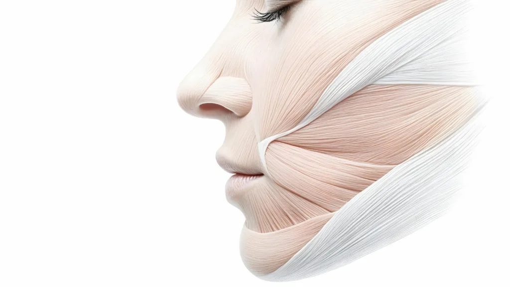
Our face is the most valuable part that makes us who we are; it is like a work of art that reflects our emotions, thoughts, and character. Understanding the structure behind this masterpiece—the facial anatomy—is the first step in grasping why and how aesthetic interventions can produce such natural results. To see our face merely as a piece of skin would be a great misconception. On the contrary, it is a complex structure, woven layer by layer, working in perfect harmony like an architectural masterpiece. The success of every aesthetic procedure depends on how well we understand this architecture and how much respect we have for it.
What Are the Anatomical Layers of Our Face?
We can think of our face as a multi-layered cake. The decoration on top—our skin—is only the visible part of the cake. The real taste and structure come from the layers beneath. In aesthetic surgery, the goal is not only to correct the decoration but to address the issues in the deeper layers that form the foundation of the cake. Only in this way can lasting and natural results be achieved. Our face is made up of five main anatomical layers, organized from the surface down to the depth. These layers are:
- Skin
- Subcutaneous Fat Tissue
- SMAS (Superficial Musculoaponeurotic System)
- Deep Fat and Connective Tissue
- Periosteum (Bone Membrane)
Now, let’s take a closer look at what these layers do. The skin is our outermost protective shield, and its quality determines how fresh and vibrant our face appears. Just beneath it lies the subcutaneous fat tissue, which contains the superficial fat pads that give our face its full, healthy, and youthful expression. The third layer, the SMAS, is the key player in modern facial aesthetics. It is an organized fibrous network that wraps around the facial muscles like a web and transmits their movements to the skin. The main culprit behind facial sagging is the loosening of this SMAS layer. The fourth layer, the deep fat and connective tissue, contains the deep fat compartments that provide the face’s fundamental volume and structural support. Most importantly, the main branches of the facial nerve travel safely through this protected corridor. At the bottom, in the fifth layer, lies the periosteum—a solid foundation that directly covers the facial bones and anchors all the soft tissues. This layered structure is very different from the rest of the body and explains why aesthetic procedures on the face require such precise anatomical knowledge.
Why Is the Anatomy of the SMAS, the Key Point of Facelift Surgeries, So Important?
Anyone who researches aesthetic surgery inevitably encounters that magical abbreviation: SMAS. This term represents a revolution in facial rejuvenation surgery. Imagine the SMAS as a strong yet flexible supportive net, lying just beneath the skin and wrapping around the entire face like a hammock. This network connects our facial muscles to one another and to the skin. When we smile, frown, or raise our eyebrows in surprise, it is this system that transmits the movement of the muscles to the skin.
So, why is SMAS anatomy so vital? Because over time, under the relentless effect of gravity, it is not the skin but the SMAS network and the soft tissues above it that sag and slide downward. In the early days of facelift surgeries, only the skin was tightened. Since this approach ignored the sagging foundation underneath, the results were neither long-lasting nor natural and often led to an undesired, artificial, “pulled” facial expression.
Modern facelift techniques, however, address the root of the problem by targeting the SMAS directly. During surgery, this supportive network is carefully released and repositioned to its more youthful, elevated location. In other words, the sagging foundation of the building is reinforced. The skin is then redraped over this solid base without any tension, with only the excess being removed. The result? As impressive as it sounds: much more natural, incredibly long-lasting, and—most importantly—a fresh appearance without the telltale signs of surgery.
Another fascinating feature of the SMAS is its continuity. It is integrated with the platysma muscle in the neck and the fascia in the temple region. This means that any intervention on the SMAS during a facelift produces a domino effect—tightening the sagging areas of the neck and jawline while simultaneously creating a subtle lift in the temple and eyebrow regions. In other words, by addressing one key structure, a comprehensive rejuvenation of the entire face can be achieved.
Why Do Facial Fats Behave Differently With Age?
One of the most striking signs of facial aging is the change in volume. While youthful cheeks appear round and full, they gradually sink over time, and unwanted bulges form along the jawline. The reason for this is that the fat tissue in our face is not one single unit. Instead, facial fat is organized into separate “compartments” composed of small fat pads of various sizes and functions. Each compartment responds differently to the aging process. These compartments fall into two main groups, each playing a different role in aging.
The characteristics of the superficial fat compartments are as follows:
- They lie directly beneath the skin
- They are more affected by gravity
- They tend to sag downward
- They deepen the nasolabial folds
- They contribute to the formation of “jowls” along the jawline
The characteristics of the deep fat compartments are:
- They are located deeper, beneath the muscles
- They provide structural support and volume to the face
- They are prone to losing volume (deflation)
- They contribute to the flattening of the cheeks and midface
- They accentuate the hollows under the eyes
The secret of the aging mechanism lies in the interaction between these two processes. The deeper, supportive fat pads gradually melt and disappear (deflation), causing the overlying superficial fat pads and skin to lose their foundation. Once they lose this support, the superficial layers succumb to gravity and sag downward (descent). Therefore, the modern philosophy of facial rejuvenation focuses not only on lifting the sagging tissues but also on restoring lost volume to the proper foundation—rebuilding the face three-dimensionally.
How Do the Retaining Ligaments That Hold Our Face in Place Work?
There is another very important anatomical structure that prevents the soft tissues of our face from sliding uncontrollably downward: the retaining ligaments. These ligaments can be thought of as strong, fibrous “steel cables” that anchor the facial soft tissue layers to the bones—the main columns of a building. They start from the periosteum (the bone membrane) deep within the face, pass upward through all the layers, and attach to the skin, effectively securing our face to the skeletal framework.
These ligaments create specific “fixation islands” in the face. The fat compartments rest securely between the walls formed by these ligaments. As we age, these retaining ligaments, like rubber bands stretched for years, lose their elasticity and strength. When the “cables” loosen, the fat pads and soft tissues they hold can no longer resist gravity and gradually begin to descend.
These ligaments are critically important in aesthetic surgery. To perform an effective facelift, the surgeon must strategically and carefully release these retaining ligaments. If these structures (for example, the zygomatic ligaments over the cheekbones or the masseteric ligaments in the cheeks) are not released, no matter how much the tissue is pulled, it cannot be effectively repositioned upward because it remains anchored to its old position. The success of the surgery depends on correctly releasing these ligaments and then securely re-suspending the SMAS layer to a new, stronger, and higher point. In filler applications, knowing the precise location of these ligaments also serves as a crucial anatomical roadmap to ensure that injections are made in the correct plane and produce natural results.
How Does the Anatomy of Facial Muscles Create Wrinkles?
The muscles in our face differ from other skeletal muscles in the body in one very important way. Instead of starting and ending on bones, they originate from a bone or deep structure and attach directly to the skin. This unique anatomy allows us to raise our eyebrows, smile, frown, and express thousands of emotions on our face.
However, this has a price. Every facial expression we make over the years causes these muscles to repeatedly contract and relax the skin. When we are young, the collagen and elastin fibers in the skin allow it to bounce back easily after each movement. As we age, the skin loses its elasticity. It can no longer fully return to its original state after each contraction, and the creases caused by these movements become permanent. These are the “dynamic wrinkles” that appear with facial expressions. Over time, as expressions repeat, they turn into “static wrinkles” that remain even when the face is at rest.
The main dynamic wrinkles and their responsible muscle groups are:
- Forehead Lines (Frontalis Muscle)
- “11” Lines Between the Eyebrows (Corrugator and Procerus Muscles)
- Crow’s Feet (Orbicularis Oculi Muscle)
- “Bunny” Lines on the Nose Bridge (Nasalis Muscle)
- Vertical “Barcode” Lines Above the Lips (Orbicularis Oris Muscle)
- Downturned Mouth Corners (Depressor Anguli Oris Muscle)
Over time, these lines become visible even when we are not making facial expressions, turning into permanent “static” wrinkles. The aesthetic balance of the face relies on a delicate interplay between these muscle groups. The position of our eyebrows, for instance, is determined by the balance between the forehead muscle that lifts them and the frowning muscles that pull them downward. When this balance is disrupted, a tired, angry, or sad expression can become permanent.
How Does Botulinum Toxin Treatment Affect Facial Muscles?
Botulinum toxin—commonly known as Botox—is a refined treatment built precisely on this knowledge of facial muscle anatomy. It works by temporarily blocking the transmission of nerve signals to the injected muscle. When the muscle no longer receives the “contract” signal from the brain, it relaxes. As the muscle relaxes, it stops pulling on the skin, allowing wrinkles to gradually smooth out and fade away.
The success of this treatment depends entirely on anatomical knowledge and aesthetic vision. The goal is never to “freeze” or erase facial expression. On the contrary, it selectively relaxes only the overactive muscles responsible for negative expressions (like anger or fatigue) and unwanted wrinkles. For example, while the frown muscles are relaxed, the muscles that lift the eyebrows are preserved. This eliminates wrinkles while giving the face a more rested, positive, and radiant expression. A skilled practitioner analyzes the face as a whole—not just erasing a line but restoring harmony among muscles to reveal a younger and fresher look. It is an art that requires precise knowledge of the location, depth, and strength of each muscle.
Why Is Knowledge of the Facial Nerve So Critically Important in Aesthetic Procedures?
The command center controlling all our facial muscles is the facial nerve. After leaving the brain, it passes through the parotid gland in front of the ear before branching like the limbs of a tree to reach different regions of the face. These branches activate the muscles of the forehead, eyes, cheeks, lips, and neck. Damage to this nerve or its branches can lead to paralysis of those muscles, causing facial asymmetry and loss of function. Therefore, knowing and protecting the facial nerve is one of the highest priorities in aesthetic surgery.
Fortunately, anatomy provides a reliable roadmap. All branches of the facial nerve travel within one of the deeper facial layers—beneath the previously mentioned SMAS layer. This is crucial information for surgeons. Procedures performed above the SMAS (in the subcutaneous plane) are extremely safe for the nerves. When operating below the SMAS, an experienced surgeon knows exactly where the nerve runs and takes meticulous care to protect it. The facial nerve, which controls our expressions, divides into five main branches:
- Temporal Branch (Forehead and Eyebrows)
- Zygomatic Branch (Around the Eyes and Cheekbones)
- Buccal Branch (Cheeks and Upper Lip)
- Marginal Mandibular Branch (Lower Lip)
- Cervical Branch (Neck)
This consistent anatomical pattern allows experienced surgeons to perform these operations with maximum safety.
What Should Be Known About Facial Danger Zones and Vascular Anatomy?
There are certain areas on the face where important nerves or blood vessels lie closer to the skin, making them more delicate during surgical or injection-based procedures. These are referred to in medicine as “danger zones.” This doesn’t mean they should be feared—it simply means professionals working in these areas must have deep anatomical knowledge and adjust their techniques accordingly.
For example, in the temple region, the nerve branch responsible for forehead movement runs superficially; careless work in this area could result in permanent eyebrow drooping. Similarly, along the jawline, the nerve that controls the muscles of the lower lip is also in a vulnerable position.
Vascular anatomy is particularly important for filler injections. There is an area known as the “danger triangle” of the face, stretching from the corners of the mouth to the bridge of the nose. The vessels here are directly connected to blood vessels inside the brain. If filler material is accidentally injected into an artery, it can block blood flow, causing tissue loss or, worse, blindness. Because the course of facial arteries varies from person to person, even “simple” procedures like fillers must be performed only by experienced physicians with thorough anatomical knowledge and the ability to manage risks. Safety must always come before aesthetics.
How Does Facial Aging Occur Step by Step?
Facial aging is not just about wrinkles or sagging skin. It is a three-dimensional process that begins at the deepest foundation and affects every layer up to the outermost skin. We can think of it as a chain of “collapse and descent.” Facial aging usually follows these stages:
- Resorption begins in the bone structure, weakening skeletal support.
- The deep fat pads above this weakened foundation lose volume (deflation).
- The retaining ligaments stretch, weaken, and lose elasticity.
- The superficial fat compartments, having lost their anchoring, sag downward due to gravity.
- The skin, now lacking support and elasticity, becomes loose and develops wrinkles.
This integrated model explains why facial rejuvenation should not focus on a single issue but instead take a holistic approach that considers every layer. The solution must always target the root cause of the problem.
How Do Modern Aesthetic Approaches Address the Anatomy of Aging?
The beauty of modern aesthetic practice lies in the fact that it offers a solution for each stage of the aging process. Instead of addressing a single issue, it treats the face as a whole, aiming to reverse every step of the “collapse and descent” sequence. Each anatomical problem has its modern aesthetic counterpart. The main problems and their solutions are as follows:
- Problem: Volume Loss (Deflation) → Solution: Fillers and Fat Injections
- Problem: Sagging (Descent) → Solution: Face and Neck Lift Surgeries
- Problem: Dynamic Wrinkles → Solution: Botulinum Toxin Applications
- Problem: Skin Quality Loss → Solution: Laser, Peeling, and Mesotherapy

Op. Dr. Erman Ak is an internationally experienced specialist known for facial, breast, and body contouring surgeries in the field of aesthetic surgery. With his natural result–oriented surgical philosophy, modern techniques, and artistic vision, he is among the leading names in aesthetic surgery in Türkiye. A graduate of Hacettepe University Faculty of Medicine, Dr. Ak completed his residency at the Istanbul University Çapa Faculty of Medicine, Department of Plastic, Reconstructive and Aesthetic Surgery.
During his training, he received advanced microsurgery education from Prof. Dr. Fu Chan Wei at the Taiwan Chang Gung Memorial Hospital and was awarded the European Aesthetic Plastic Surgery Qualification by the European Board of Plastic Surgery (EBOPRAS). He also conducted advanced studies on facial and breast aesthetics as an ISAPS fellow at the Villa Bella Clinic (Italy) with Prof. Dr. Giovanni and Chiara Botti.
Op. Dr. Erman Ak approaches aesthetic surgery as a personalized art, tailoring each patient’s treatment according to facial proportions, skin structure, and natural aesthetic harmony. His expertise includes deep-plane face and neck lift, lip lift, buccal fat removal (bichectomy), breast augmentation and lifting, abdominoplasty, liposuction, BBL, and mommy makeover. He currently provides safe, natural, and holistic aesthetic treatments using modern techniques in his private clinic in Istanbul.


